 .
.
You will notice from the intensity profiles in the previous section
that there are fewer samples in the z dimension than in the x and y
dimensions. The clinical MR volumes are acquired as a sequence of
slices in the axial (z) direction. The result is a sequence of images
such as those shown in Figure 2.3. In this case, the
data set consists of 27 5mm thick slices; the pixel dimensions in each
slice are  .
.
The images displayed in Figure 2.3 represent the average of the FID response signals from all tissues in the 5mm slice. Thus, each voxel in the images could represent more than one tissue type. This phenomenon is referred to as the partial volume effect.
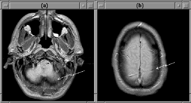
The partial volume effect is particularly noticeable in the extreme slices of MRI volumes. Figure 2.6 contains the first and last slice of a PD-weighted MRI data set. Brain tissue in the lower right of the first slice appears dim because tissues below the brain (with smaller FID response signals) contribute to the intensity of the slice. The boundary of the brain in the last slice is blurred because the voxels in that area represent both brain and cerebral spinal fluid (CSF). Obviously, the partial volume effect can hinder the detection of the intracranial boundary.

Figure 2.6: The partial volume effect
results in dim brain tissue in the first MRI slice (a) and blurs the
edge of the brain in the last slice (b).