In MR images of the head, we can assume that any voxel that is not background noise must be part of the head. Therefore, if we can determine what the background noise looks like, we can isolate the head. The problem of locating the intracranial boundary then becomes simpler because we know (approximately) where, within the head, the brain should be.
By assuming the MR scanner produces normally distributed white noise, Brummer et al. have shown that background noise in the reconstructed PD-weighted MR volumes has a Rayleigh distribution [7]:

where  is the noise intensity and
  is the standard
deviation of the white noise. As shown in
Figure 2.7, this distribution is easily visible
in the low intensity range of the uncorrected MRI histogram.
is the standard
deviation of the white noise. As shown in
Figure 2.7, this distribution is easily visible
in the low intensity range of the uncorrected MRI histogram.
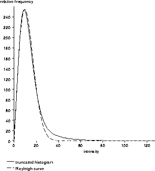
Figure 2.7: A truncated histogram of a
PD-weighted MR volume. The background noise at the low end of the
histogram is characterized by a Rayleigh distribution.