 ,
with standard deviation,
  :
:
Brummer et al. also characterize brain tissue intensities in
uncorrected, PD-weighted MR scans [7]. They show that
voxels inside the brain have normally distributed intensities,  ,
with standard deviation,
  :
:

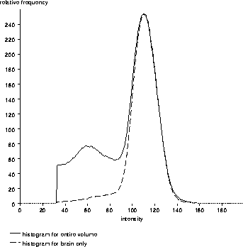
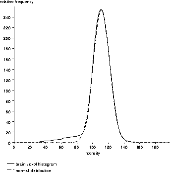
Figure 2.8 shows the histogram of brain voxel intensities superimposed on the high-intensity part of a PD-weighted MRI histogram. Indeed, the distribution of brain voxel intensities appears to be normally distributed. Qualitatively, Figure 2.9 verifies that the histogram of brain voxel intensities represents a normal distribution.

Figure 2.8: The high-intensity
region of a histogram of a PD-weighted MR volume with the brain voxel
histogram overlaid. The intensities of the brain voxels appear to be
normally distributed.

Figure 2.9: Overlaying a
normal distribution on the brain voxel intensity histogram shows that
the brain voxel intensities are (approximately) normally distributed
in a PD-weighted MRI volume.
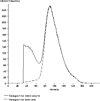
Brain voxel intensities in a T2-weighted MR scan are somewhat normally distributed. However, Figure 2.10 shows that the distribution of brain voxel intensities is obviously skewed.

Figure 2.10: The high-intensity
region of a histogram of a T2-weighted MR volume with the brain voxel
histogram overlaid. The intensities of the brain voxels are not quite
normally distributed.