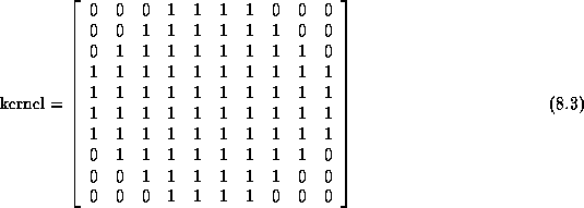
Results for the Generate Initial Brain Mask process are presented and discussed in this section. The process igraph uses similar parameters to produce initial brain masks for the data sets acquired by each scanner. These parameters are listed in Tables 8.5. Only the fieldOfView and sliceGap parameters of the mriBlurVolume operator differ to correspond with MRI acquisition parameters. In all cases, the kernel used by berode and bdilate is the following binary matrix:
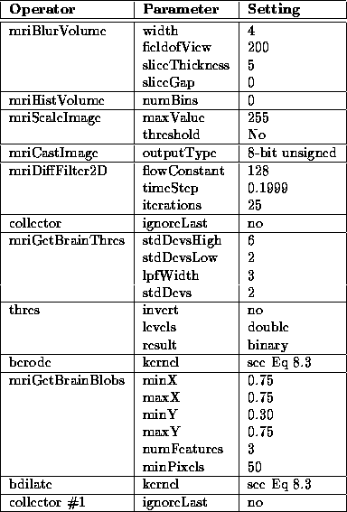
Table 8.5: Generate Initial
Brain Mask igraph operator parameters used for Data Set 1
The initial brain mask for Data Set 1, produced by the Generate Initial Brain Mask process, is overlaid on the PD-weighted MR volume in Figure 8.11. Slice 7 is enlarged in Figure 8.12.
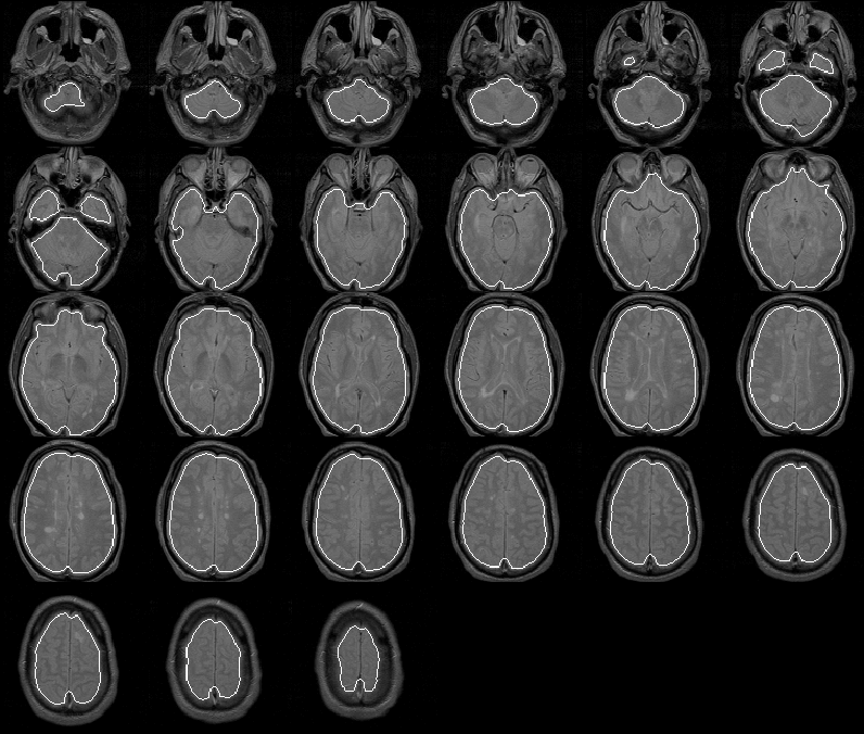
Figure 8.11: The initial brain mask for
MRI Data Set 1 overlaid on the PD-weighted scan.
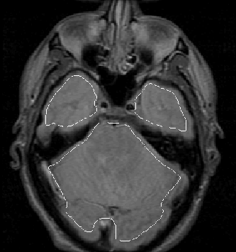
Figure 8.12: The initial brain mask
for slice 7 of MRI Data Set 1 overlaid on the PD-weighted image.
The mask apparently identifies all brain tissue regions. Its boundary falls consistently close to, and inside, the intracranial boundary. The initial brain mask should be an ideal seed for the Generate Final Brain Mask process which uses the active contour model algorithm with balloon forces to push the mask boundary outward.
As shown in Figure 8.13, the Generate Initial Brain Mask produces an initial brain mask for Data Set 2 that is comparable to that produced for Data Set 1. The brain mask provides a good seed for the Generate Final Brain Mask process.
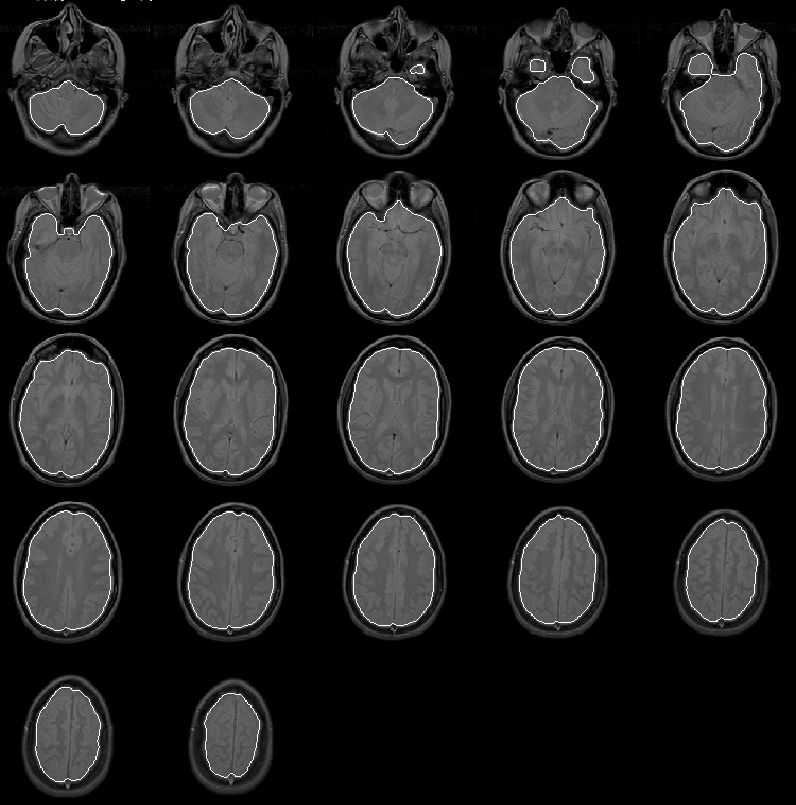
Figure 8.13: The initial brain mask for
MRI Data Set 2 overlaid on the PD-weighted scan.
For Data Set 3 the Generate Initial Brain Mask process again produces a good initial brain mask to use as a seed for the Generate Final Brain Mask process. The mask is overlaid on the PD-weighted scan of Data Set 3 in Figure 8.14.
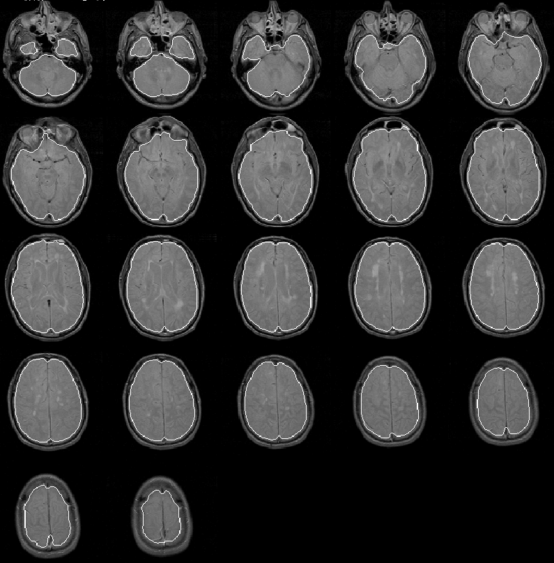
Figure 8.14: The initial brain mask for
MRI Data Set 3 overlaid on the PD-weighted scan.
Figure 8.15 shows the initial brain mask for Data Set 4 overlaid on the PD-weighted scan. As with the previous three data sets, the mask is very good. In slices 11 through 14, the mask extends beyond the intracranial boundary due to the bright areas between the eyes and in the forehead region of the patient. Still, the mask is close enough to the intracranial boundary for the Generate Final Brain Mask process to work effectively.
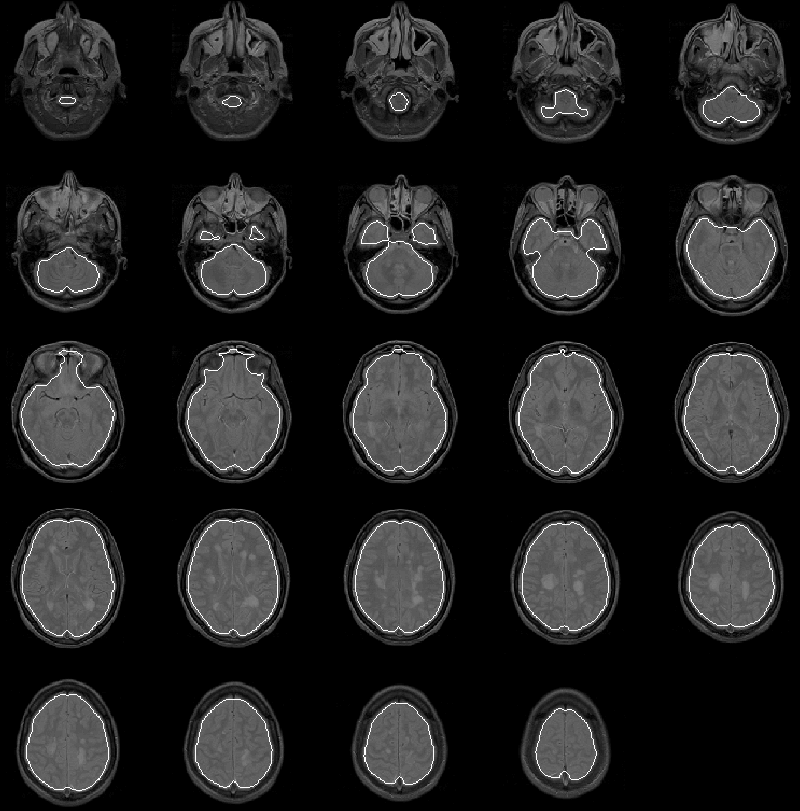
Figure 8.15: The initial brain mask for
MRI Data Set 4 overlaid on the PD-weighted scan.
The initial brain mask for Data Set 5 is overlaid on the PD-weighted scan in Figure 8.16. The mask is, again, very good. Only a small region of extremely low intensity brain voxels is missed in the lower left corner of slice 2.
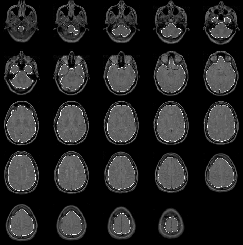
Figure 8.16: The initial brain mask for
MRI Data Set 5 overlaid on the PD-weighted scan.
Approximate benchmarks for the Generate Initial Brain Mask igraph are listed in Table 8.6 for the 5 data sets. The benchmarks were recorded for the igraph running on a Sun SPARC 5 workstation with 16 MBytes of RAM.
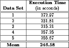
Table 8.6: Approximate
benchmarks for the Generate Initial Brain Mask igraph running an
a SUN SPARC 5 workstation.
The Generate Initial Brain Mask process detects the brain for every data set tested herein. The mean execution time of the process running on a Sun SPARC 5 workstation was 4.11 minutes. The resulting initial brain masks contain no significant misclassified regions. The masks provide excellent seeds for the Generate Final Brain Mask process.