
The outcome of the Segment Head process for each of the 5 MRI data sets is investigated in this section. The head mask for each data set is presented and its validity is discussed.
The operator parameters for the Segment Head igraph remain constant for all the data sets. These parameters are listed in Table 8.3. The kernel used by berode and bdilate is the following binary matrix:
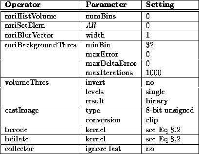
Table 8.3: Segment Head igraph operator
parameters.
The boundary of the head mask produced for Data Set 1 is overlaid on the PD-weighted volume in Figure 8.6. For the most part, the mask is extremely accurate.
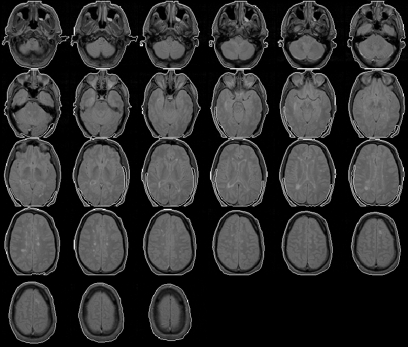
Figure 8.6: The head mask for MRI Data Set 1
overlaid on the PD-weighted scan.
In some slices, the mask does not cover the entire head. This anomaly occurs because the mriFillBlobs operator in the Segment Head igraph fails to fill in regions that are not completely enclosed, even at the image edges. The problem might be fixed by treating the image edge as an enclosing boundary.
This problem has not been addressed because it has little effect on the final results of the intracranial boundary detection algorithm. The Generate Initial Brain Mask process uses the centroid and bounding box of the head mask in each slice. The inaccuracies in the mask affect these features only slightly.
Figure 8.7 overlays the head mask produced for MRI Data Set 2 on the PD-weighted volume. The mask appears extremely accurate except for a region in slice 6 where the it extends outside the head. This error is caused by the of presence falsely placed data voxels due to phase-encoding problems in MR image reconstruction (see Chapter 2). This error might be reduced by low pass filtering or median filtering the volume before thresholding. As with the error occurring in Data Set 1, this error has little impact on the final results of the intracranial boundary detection algorithm and therefore has not been addressed.
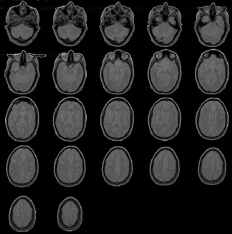
Figure 8.7: The head mask for MRI Data Set 2
overlaid on the PD-weighted scan.
Figure 8.8 shows that the head mask produced for Data Set 3 is comparable to those produced for Data Sets 1 and 2. Here, the head mask is relatively error-free.
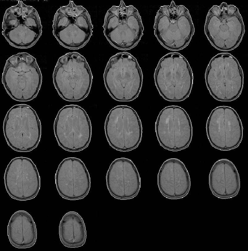
Figure 8.8: The head mask for MRI Data Set 3
overlaid on the PD-weighted scan.
The Segment Head process produces a head mask for Data Set 4 with no significant errors. The head mask is overlaid on the PD-weighted volume in Figure 8.9.
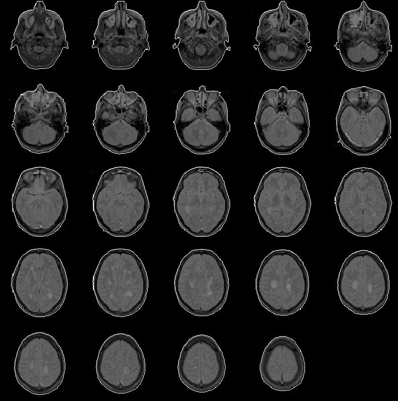
Figure 8.9: The head mask for MRI Data Set 4
overlaid on the PD-weighted scan.
The head mask for Data Set 5 is overlaid on the PD-weighted volume in Figure 8.10. The mask contains minor errors similar to those in the head mask of Data Set 2.
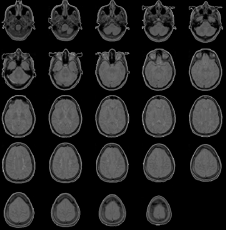
Figure 8.10: The head mask for MRI Data Set 5
overlaid on the PD-weighted scan.
The approximate execution time for the Segment Head igraph is listed in Table 8.4 for each of the 5 data sets. These benchmarks were recorded for the igraph running on a Sun SPARC 5 workstation with 16 MBytes of RAM.
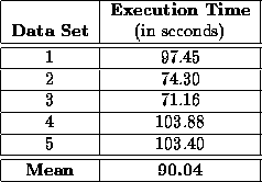
Table 8.4: Approximate benchmarks for the Segment Head igraph running an a SUN SPARC 5 workstation.
The Segment Head process produces a reasonable head mask for each of the 5 data sets in an average time of 1.5 minutes. Some of the masks contain minor errors that will not inhibit the Generate Final Brain Mask process. The errors are probably correctable but, because the Generate Initial Brain Mask process tolerates the errors, their removal is left for future work.