Each of the 5 MRI data sets studied herein were acquired using dual echo spin-echo sequences. In such sequences, PD-weighted and T2-weighted image modalities are acquired simultaneously and, therefore, do not require registration. See Chapter 2 for details.
The first three data sets were acquired using a General Electric 1.5 Tesla MRI scanner at London, Ontario. The last two were acquired using a 1.5 Tesla MRI scanner at U.B.C. Hospital. The acquisition parameters for each of the bimodal MRI data sets are listed in the following sections.
Data Set 1 has been used in experiments throughout this
manuscript. Its acquisition parameters are listed in
Table 8.1. The original 12-bit data have been scaled to
8 bits by the MRI lab technician. Each original voxel intensity,
 , is scaled to
  according to the maximum
intensity,
according to the maximum
intensity,  , in the volume:
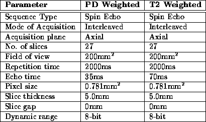
Table 8.1: MRI Acquisition Parameters for Data Set 1
Figure 8.1 shows three slices selected from Data Set 1. The extreme slices (particularly the lowest slice) exhibit considerable partial volume effect resulting in a non-distinct intracranial boundary. The boundary is difficult to detect accurately in these slices. Except for intensity variation due to RF inhomogeneity, other slices in the volume contain no remarkable features.
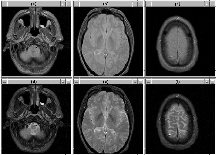
Figure 8.1: Selected slices from MRI Data Set 1. (Top)
PD-weighted. (Bottom) T2-weighted. (a), (d) Slice 1. (b), (e)
Slice 14. (c), (f) Slice 27.
The acquisition parameters for Data Set 2 and Data Set 3
are similar to those for Data Set 1, except that the field of view is
210mm (pixel size = 0.820mm ) and that the data sets contain 22
slices. Similarly, the data have been scaled according to
Equation 8.1.
) and that the data sets contain 22
slices. Similarly, the data have been scaled according to
Equation 8.1.
Selected slices for Data Set 2 are shown in Figure 8.2. Selected slices for Data Set 3 are shown in Figure 8.3. Notice that there is a region of bright voxels in slice 9, just above the brain, that might affect the detection of the intracranial boundary.
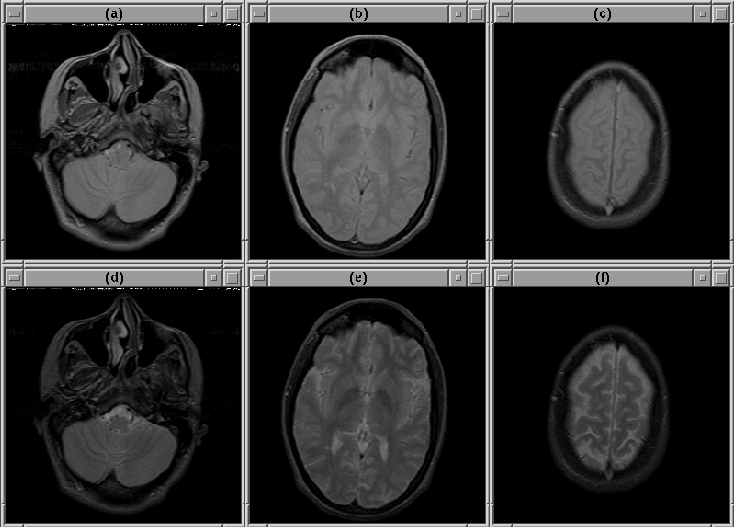
Figure 8.2: Selected slices from MRI Data Set 2. (Top)
PD-weighted. (Bottom) T2-weighted. (a), (d) Slice 1. (b), (e)
Slice 11. (c), (f) Slice 22.
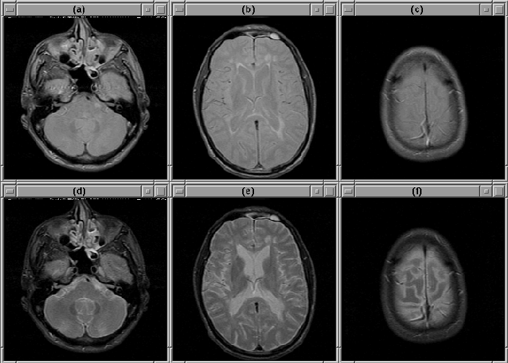
Figure 8.3: Selected slices from MRI Data Set 3. (Top)
PD-weighted. (Bottom) T2-weighted. (a), (d) Slice 1. (b), (e)
Slice 9. (c), (f) Slice 22.
Data Set 4 and Data Set 5 were acquired using a newer MRI scanner than the previous three data sets. The images have not been scaled. The acquisition parameters for these data sets are listed in Table 8.2.
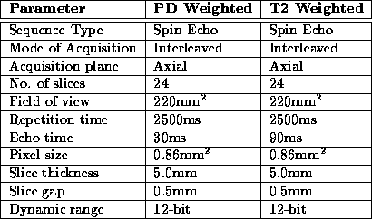
Table 8.2: MRI Acquisition Parameters for Data Sets 4 and 5
The data sets appear to be of higher quality than the previous data sets. This perception is probably due to the fact that the data sets have a 12-bit dynamic range as opposed to an 8-bit dynamic range; in fact, the resolution of Data Sets 4 and 5 is lower than that of the previous data sets.
Figure 8.4 shows slices selected from Data Set 4. The lower slices contain bright anomalies, that might be due to clogged sinuses or dental work, in regions outside the brain. These anomilies could confuse the intracranial boundary detection algorithm. Other bright anomilies, perhaps fat deposits, appear in the forehead area of the patient (see slice 12).
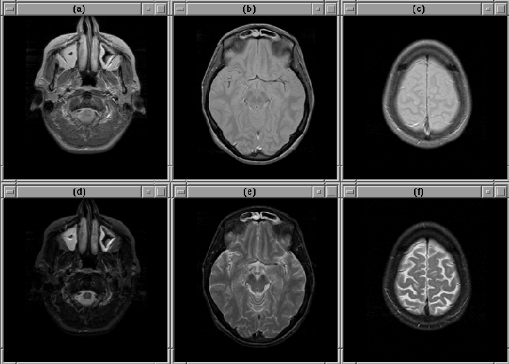
Figure 8.4: Selected slices from MRI Data Set 4. (Top)
PD-weighted. (Bottom) T2-weighted. (a), (d) Slice 2. (b), (e)
Slice 12. (c), (f) Slice 24.
Figure 8.5 shows selected image slices from Data Set 5. Except for some obvious partial volume effects in slice 2, there are no remarkable features in the volume.
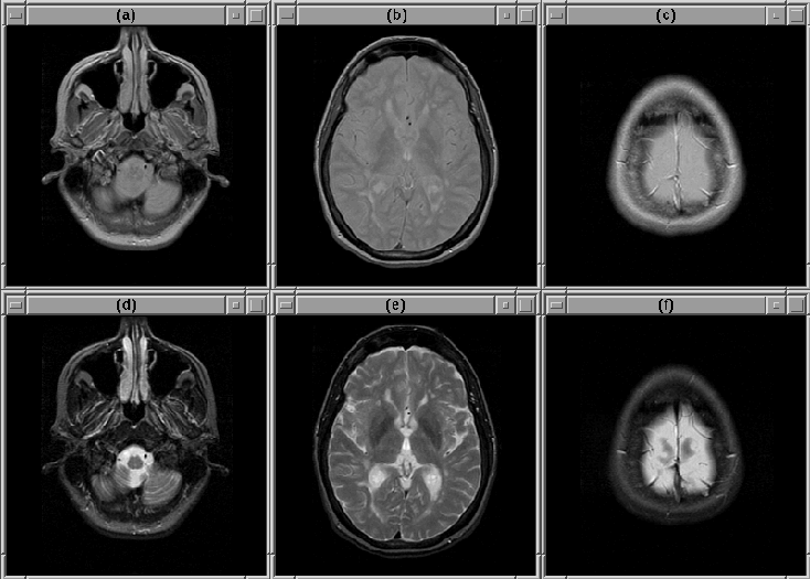
Figure 8.5: Selected slices from MRI Data Set 5. (Top)
PD-weighted. (Bottom) T2-weighted. (a), (d) Slice 2. (b), (e)
Slice 12. (c), (f) Slice 24.
Data Sets 4 and 5 exhibit very little intensity variation due to RF inhomogeneity. RF correction would not likely improve image quality.
