Note that the initial brain mask is imprecise. The active contour model algorithm, presented in Chapter 5, is used to refine the initial brain mask in a completely automatic process. The igraph implementing the process, Generate Final Brain Mask is shown in Figure 7.14. Brief descriptions of the igraph's operators are provided in Table 7.5. The igraph is discussed in more detail in the following sections.
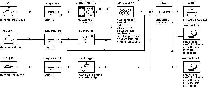
Figure 7.14: WiT igraph implementing the
Generate Final Brain Mask process.
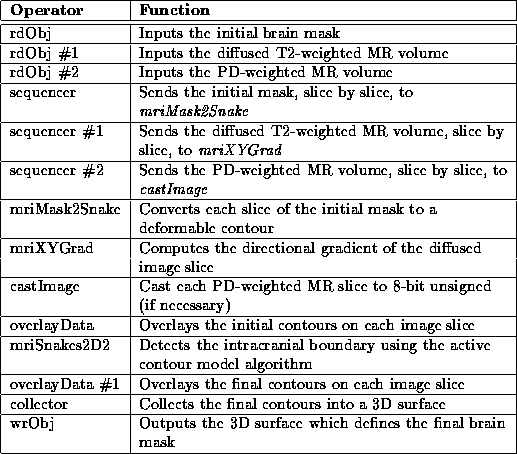
Table 7.5: A brief
description of the operators used in the Generate Final Brain
Mask igraph.
rdObj inputs the initial brain mask which is sent, slice by slice, to mriMask2Snake. mriMask2Snake converts each mask slice to a deformable contour for input into mriSnakes2D2. The igraph for mriMask2Snake is given in Figure 7.15. Its operator descriptions are listed in Table 7.6.
Figure 7.15: WiT hierarchical
igraph implementing mriMask2Snake.
Table 7.6: A brief
description of the operators used in the mriMask2Snake
hierarchical igraph.
mriMask2Graphic converts each independent region in the mask slice to a deformable contour by tracing the boundary of the region. However, the points in the resulting contour correspond to adjacent pixels which will cause instability in the active contour model algorithm (see Chapter 5). Therefore, we resample the contour to increase the distance between adjacent points. This resampling is achieved by mriResizeGraphic.
The contour is decimated by a factor of 6 to a minimum of 16 points. This factor was chosen experimentally to produce good results for all data sets.
Referring back to Figure 7.14, mriXYGrad computes the directional gradient of each slice in the diffused MR volume produced by the Generate Initial Brain Mask process. As illustrated in Figure 7.16, mriXYGrad is a hierarchical igraph that uses standard WiT operators.

Figure 7.16: WiT hierarchical igraph
implementing mriXYGrad.
Given initial contours, mriSnakes2D2 uses the active contour model algorithm described in Chapter 5 to detect the intracranial boundary. It uses diffused T2-weighted slices for gradient information because they contain strong, gradual edges that stabilize the algorithm. PD-weighted slices are used for intensity information because, in these slices, tissue intensities inside the brain are relatively homogeneous and distinct from the surrounding tissue intensities. These characteristics inhibit the deformable contours from attaching themselves to physiological features within the brain and move the contours closer to the intracranial boundary.
Figure 7.17 shows the refinement of the initial contours by the active contour model algorithm for slice 6 of Data Set 1. Complete results are presented in Chapter 8.
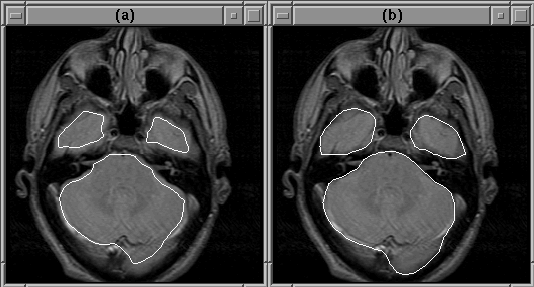
Figure 7.17: Refinement of the brain mask by
the active contour model algorithm (a) Initial contour. (b) Final
contour.