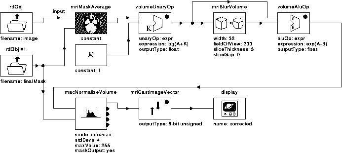
Figure 7.18: WiT igraph implementing the Correct Intensity process.
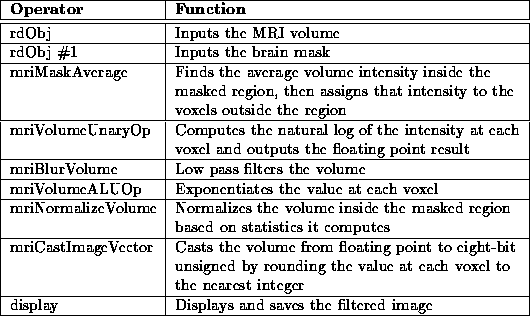
The Correct Intensity process uses the form of homomorphic filtering presented in Chapter 6 to reduce intensity variation due to RF inhomogeneity in MR image volumes. The WiT igraph for the process is given in Figure 7.18. The igraph operators are summarized in Table ~7.7.

Figure 7.18: WiT igraph implementing the Correct Intensity process.

Table 7.7: A brief description of
the operators used in the Correct Intensity igraph.
The igraph consists of three processing steps:
These steps are described in the following sections.
The large contrast between data voxels, such as the brain, eyes, skin, fat, etc., and background voxels contributes significantly to the low frequency spectrum of the MRI volumes. The homomorphic filter attenuates these low frequency components, erroneously attributing this contrast to RF inhomogeneity. Therefore, the contrast must be reduced. Because we are only interested in tissues within the brain, mriMaskAverage realizes the contrast reduction by assigning voxel intensities similar to those found within the brain to all voxels outside the intracranial boundary.
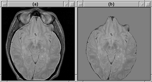
rdObj #1 inputs the brain mask produced by the Generate Final Brain Mask process and sends it to mriMaskAverage. Using the mask, mriMaskAverage finds the average intensity of brain voxels in the MR volume input by rdObj. It then assigns the average intensity to voxels outside the mask. Figure 7.19 shows the result of the masking operation.

Figure 7.19: An MR image slice (a) before masking
and (b) after. The intracranial boundary is overlaid on (a).
The three operators, volumeUnaryOp, mriBlurVolume, and
volumeALUOp, homomorphic filter the masked volume. volumeUnaryOp computes the natural logarithm of the intensity at each
voxel. An offset of 1 is added to the intensity of each voxel so that
 is never computed. mriBlurVolume
(approximately) low pass filters the log-domain volume as described in
Section 6.4. It accounts for the anisotropicity of the
volume. volumeALUOp subtracts the low pass filtered volume from
the original, then exponentiates the result.
volumeALUOp outputs an intensity-corrected, floating point volume. The mean voxel intensity of the volume is zero as a result of the homomorphic filtering operation. The segmentation algorithm (see Section 1.1), for which intensity correction is necessary, requires 8-bit unsigned volumes for input. Therefore, the corrected MR volume must be normalized and cast appropriately.
mriNormalizeVolume finds the maximum and minimum intensity of voxels within the intracranial boundary (defined by the final brain mask). It then scales each voxel within the boundary as follows:

where  and
and  are the original and scaled voxel
intensities respectively, and
  and
and  are the
maximum and minimum voxel intensities. The intensities of voxels
outside the intracranial boundary are set to zero.
mriCastImageVector casts the scaled floating point volume to 8-bit unsigned by rounding each voxel intensity to the nearest integer. display displays and saves the corrected volume.
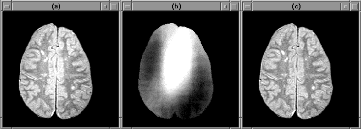
Figure 7.20 shows slice 19 of the PD-weighted Data Set 1 before and after intensity correction. Results, including quantitative measures for several data sets are presented in Chapter 8.

Figure 7.20: An MR image slice (a) before
intensity correction and (c) after. (b) shows the RF inhomogeneity.