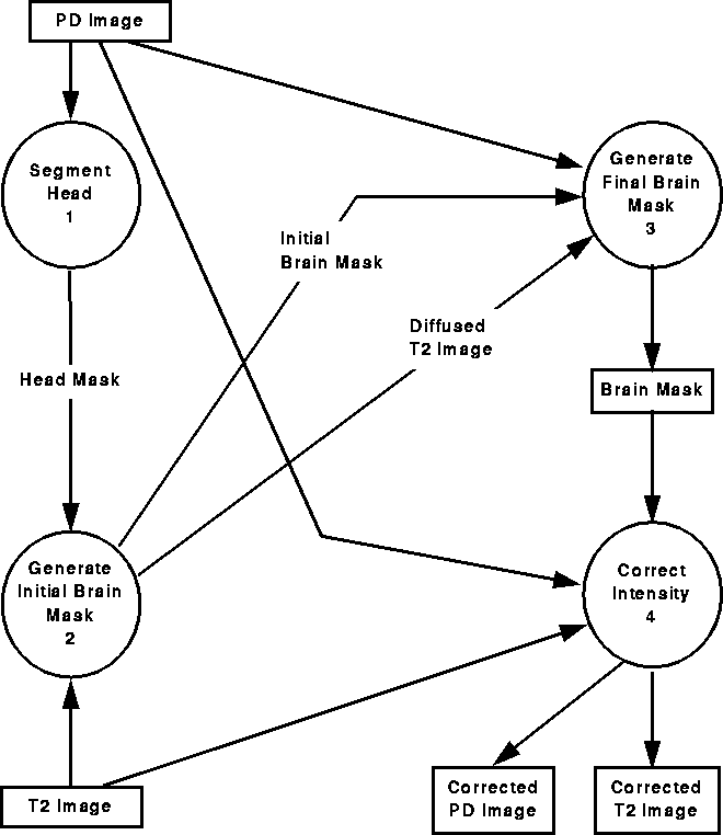
Figure 7.1: A simplified data flow diagram representing automatic intracranial boundary detection and RF correction.
Recall from Chapter 1 that the primary goal of this thesis is to develop a preprocessing step that will improve the performance MS lesion segmentation algorithms such as Johnston's [22]. Because these algorithms assume that similar tissue types have similar intensities everywhere in MRI scans, they produce poor results in the presence of intensity variation due to RF inhomogeneity. Segmentation algorithms also have difficulty dealing with tissues outside the brain, such as skin and fat, because these tissues can have similar intensities as brain tissues and MS lesions. Consequently, intensity correction and the removal of tissues outside the intracranial boundary are necessary for successful segmentation.
We have developed a novel fully automatic intracranial boundary detection and RF correction method consisting of four steps:
A simplified data flow diagram representing the method is illustrated in Figure 7.1. Each step (bubble) in the diagram has been implemented using the WiT visual programming environment introduced in Chapter 1. These steps, which execute in the order of their labels, are summarized below.

Figure 7.1: A
simplified data flow diagram representing automatic intracranial
boundary detection and RF correction.
The method assumes that PD-weighted and a T2-weighted MRI data sets have been simultaneously acquired. Using histogram analysis of the PD-weighted data set, the first step, Segment Head, isolates the area within the head and provides a head mask defining that region. The second step, Generate Initial Brain Mask, produces a mask that approximately identifies the intracranial boundary. It uses nonlinear anisotropic diffusion of the T2-weighted data set and spatial information provided by the head mask to remove regions outside the brain. With the initial brain mask as a seed, the third step, Generate Final Brain Mask, locates the intracranial boundary using an active contour model algorithm. Finally, the Correct Intensity step reduces intensity variation (due to RF inhomogeneity) within the detected intracranial boundary by homomorphic filtering the MR volume under the final brain masks.
The four steps are explained in detail in the following sections. We include example results for one MRI data set in order to clarify each step. Results for several data sets are presented in Chapter 8.