"Probabilistic models in Automated Skin lesion diagnosis"


Skin Lesion Segmentation using the Random Walker Method

 Abstract:

We present a method for automatically segmenting skin lesions by initializing the random walker algorithm with seed points whose properties, such as colour and texture, have been learnt via a training set. We leverage the speed and robustness of the random walker algorithm and augment it into a fully automatic method by using supervised statistical pattern recognition techniques. We validate our results by comparing the resulting segmentations to the manual segmentations of an expert over 120 cases, including 100 cases which are categorized as difficult (i.e.: low contrast, heavily occluded, etc.). We achieve an F-measure of 0.95 when segmenting easy cases, and an F-measure of 0.85 when segmenting difficult cases.

Introduction

  • We have developed a fully automatic skin lesion segmentation method by leveraging texture metrics, a supervised learning, and the Random Walker segmentation algorithm.

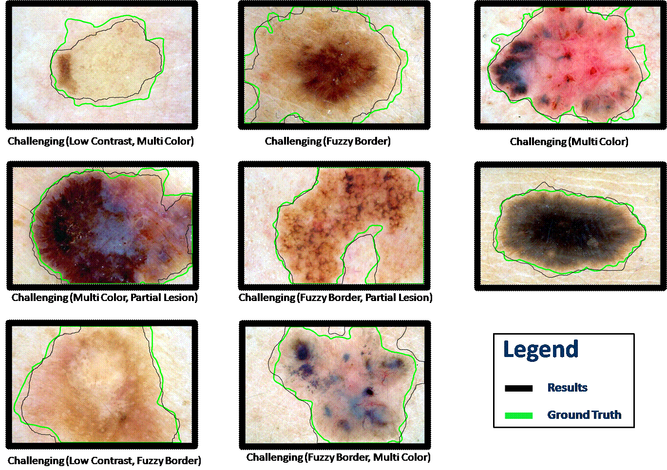
  • We validate our method using a challenging set of images where:
    1) Contrast between skin and lesion is low
    2) Lesion border is not clearly defined (Fuzzy Border)
    3) The entire border is not visible in the lesion
    4) There is considerable occlusion (hair or oil)
    5) There are many different colours present

Method


  • Texture features are created by convolving the images with a filterbank consisting of Gaussian and Laplacian of Gaussian filters.


  • Using expertly labeled ground truth and Linear Discriminant Analysis (LDA) the optimal linear combination of texture features which separates the groups of pixels (into lesion/background) is determined. The probability that each pixel belongs to the lesion is computed.

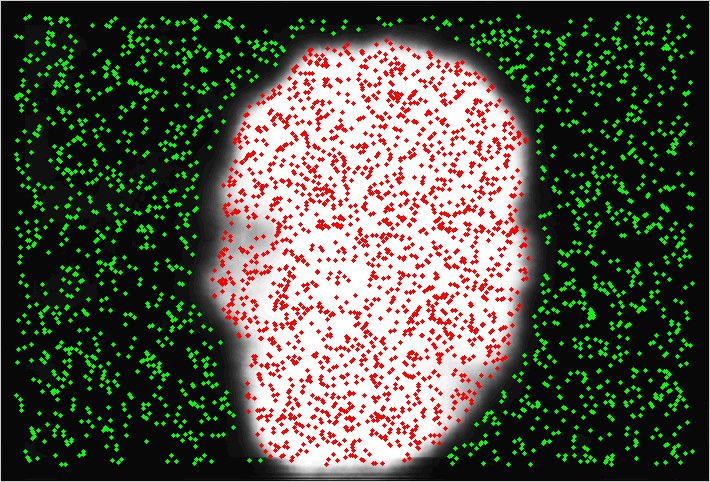
  • A histogram analysis of these ‘probability images’ determines candidate seed points. We fit a Gaussian Mixture Model to the histogram and extract the dominant Gaussians that represent the certain skin and lesion boundaries.

  • Seed points are placed and the random walker algorithm [1] is used to segment the lesion.

  • For the uncertain values Random Walker method labels the pixels.


Results

Publication (PDF) (Poster): P. Wighton, M. Sadeghi, T.K. Lee and M.S. Atkins. “A fully automatic random walker segmentation for skin lesions in a supervised setting.”  Medical Image Computing and Computer Assisted Interventions – MICCAI 2009, Springer-Verlag Lecture Notes in Computer Science, Sept. 2009, London, UK.