Dermoscope Calibration
We are working on a method to calibrate inexpensive digital dermoscopes, made by attaching a traditional dermoscope to a consumer grade digital camera. Such ‘low-cost’ digital dermoscopes are becoming increasingly popular as the prevalence of dermoscopic use in the clinic increases, and the cost of digital cameras decreases.
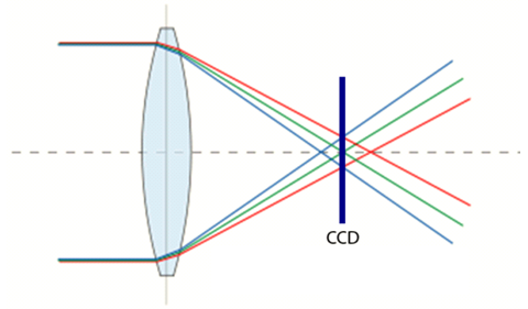
In addition to calibrating for color and lighting, we examine a distortion effect known as chromatic aberration, and correct for it accordingly. Chromatic aberration occurs when the refractive index of the optics is not constant with respect to wavelength. While distortions due to chromatic aberration can be partially alleviated by decreasing the camera aperture size (thereby restricting the use of the lens to the center where the distortion effects are minimized), such distortions are still evident when employing inexpensive components. Such distortions are visually distracting and possibly clinically confounding. Furthermore these distortions could also mislead automated characterization techniques (such as the automated identification of occluding hair or dermoscopic structures). For example, the variance of color within a lesion is an extremely important feature in both clinical as well as automated diagnosis; the accuracy of which would certainly be curbed by the introduction of spurious shades of red and blue. As a result, a software-based method of chromatic aberration correction is becoming increasingly necessary.
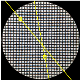
Calibration for chromatic aberration is achieved my imaging a checkered pattern and estimating the radial distortion of the red and blue channels relative to the green channel.

Before calibration, the color channels are separated causing strong shades of red and blue along edges.

After calibration, the separation of the channels is reduced considerably.

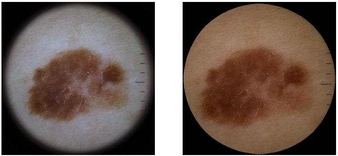
The full calibration procedure (color + lighting + chromatic aberration) applied to a dermoscopic image of a melanoma in situ

